

Turn on cookies to sign in
Signing in saves your progress to your Curie account. We can only do that with cookies on — turn them on to continue.
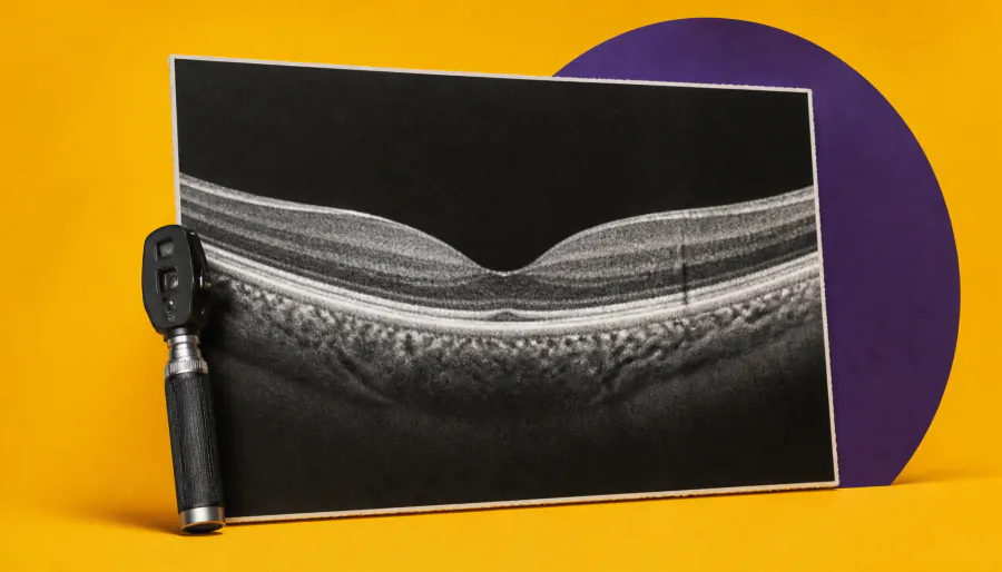
A routine OCT eye scan could do more than take a picture — it might predict the future. Researchers found that greater outer retinal volume on standard OCT imaging was linked to significantly higher odds of diabetic retinopathy (DR) progression over two years. The finding positions OCT as a potentially accessible, low-cost tool for early risk stratification in diabetic patients.
Two new studies are highlighting how retinal imaging — both standard OCT and the more advanced OCT angiography (OCT-A) — could transform how clinicians track and predict diabetic retinopathy (DR) progression.
In the primary study, researchers at Northwestern University analyzed 190 eyes from 121 diabetic patients and found that greater outer retinal volume on standard OCT was significantly associated with higher odds of DR progression over two years. Crucially, this uses widely available, lower-cost technology — unlike OCT-A, which remains expensive and not universally adopted in primary eye care. A complementary study from Vanderbilt University reinforced the value of OCT-A, showing small but consistent longitudinal changes in microvascular metrics — like vessel density and acircularity index — that tracked with DR severity over three years of repeated imaging.
Key Takeaways:
Why it matters: Diabetic retinopathy is a leading cause of preventable blindness worldwide. These findings suggest that routine OCT scans — already common in eye care settings — could be repurposed as early warning tools, helping clinicians identify high-risk patients sooner and intervene before vision-threatening complications develop.