
Turn on cookies to sign in
Signing in saves your progress to your Curie account. We can only do that with cookies on — turn them on to continue.
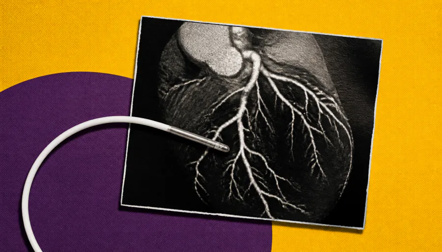
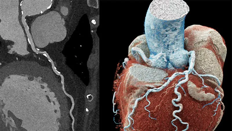
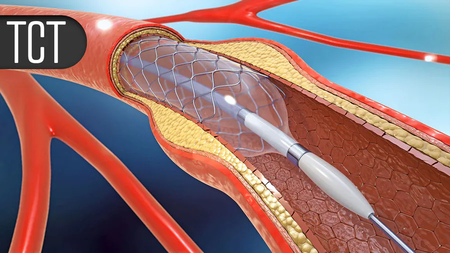
Planning heart stent procedures with CT scans ahead of time is just as effective as using ultrasound guidance during the procedure, according to the landmark P4 trial. The study found that CT-guided PCI matched IVUS-guided PCI on 12-month outcomes — and was faster and used less contrast dye. Experts say this could reshape how cardiologists plan and perform coronary interventions.
The P4 trial, presented at TCT 2026 and published in the New England Journal of Medicine, found that planning and guiding percutaneous coronary intervention (PCI) using coronary CT angiography (CCTA) was noninferior to the current gold standard — intravascular ultrasound (IVUS) guidance — for 12-month target vessel failure. The trial enrolled 1,105 patients with chronic coronary syndrome or stabilized non-ST elevation ACS across 23 international centers.
Beyond matching outcomes, the CT-guided approach offered practical advantages: procedures were about 17 minutes shorter and used 20 mL less contrast dye. The idea is to shift PCI planning upstream — letting cardiologists review detailed 3D coronary anatomy, calcium burden, and CT-derived fractional flow reserve before entering the cath lab, reducing real-time cognitive load and enabling shared decision-making with patients.
By the Numbers
Why it matters: CT-guided PCI could make heart procedures safer, faster, and less resource-intensive — but wider adoption will require better software, advanced CT scanners, and cardiologist training. A follow-up trial (P5) is already underway in higher-complexity patients.