Curie Brief
Turn on cookies to sign in
Signing in saves your progress to your Curie account. We can only do that with cookies on — turn them on to continue.
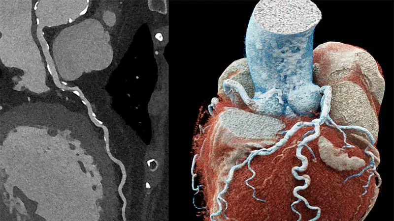
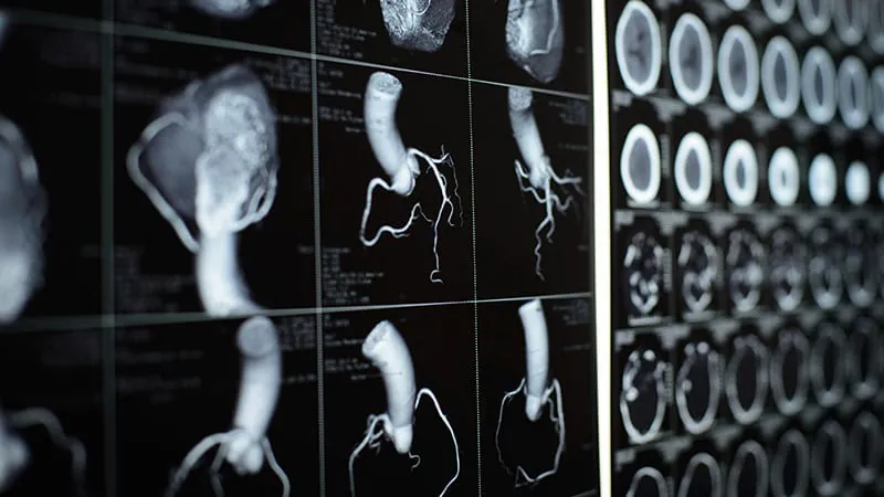
A sharper look at the heart: Photon-counting CT (PCCT) scanners are transforming cardiac imaging by converting X-ray photons directly into electrical signals, eliminating the blurring artifacts that plague conventional CT. Studies show PCCT downclassifies stenosis severity in over a third of vessels and reduces invasive coronary angiography referrals — meaning fewer unnecessary procedures and better patient outcomes. Experts say it could become the gold standard within a decade or two.
A sharper look at the heart: Photon-counting CT is changing the game in cardiac imaging
Photon-counting CT (PCCT) scanners are giving cardiologists and radiologists an unprecedented view of the heart. Unlike conventional CT machines — which convert X-rays to light and then to electrical signals, losing detail along the way — PCCT counts individual photons and converts them directly, significantly reducing image artifacts like "blooming" and blurring. The result: clearer vessel boundaries, visible high-risk fatty plaque hidden behind calcification, and tissue slices as thin as 0.2 mm (vs. 0.6 mm for conventional CT). Patients previously deemed "unscannable" — those with stents, dense calcium, or implanted devices — are now viable candidates.
The clinical payoff is significant. Multiple studies show PCCT reduces overdiagnosis and unnecessary downstream procedures, including invasive coronary angiography (CAG). Experts believe updated clinical guidelines may follow as evidence continues to mount.
By the Numbers
Why it matters: PCCT has the potential to reduce unnecessary invasive procedures, improve diagnostic accuracy for high-risk patients, and ultimately lower healthcare costs — though its $5M price tag remains a significant barrier to widespread adoption.