
Turn on cookies to sign in
Signing in saves your progress to your Curie account. We can only do that with cookies on — turn them on to continue.
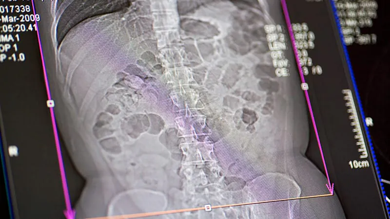
Your everyday abdominal CT might be doing double duty. A new study found that opportunistic CT scans can reliably map age-related bone density loss across the pelvis, sacrum, and lower spine — with the sharpest declines in trabecular-rich regions. Researchers say these scans could serve as an adjunctive screening signal for osteoporosis, though they're not a replacement for DEXA or FRAX assessments.
Routine abdominal CT scans may be hiding a valuable bonus: a window into bone health. A retrospective study of 330 opportunistic CT scans from adults aged 20–91 found that older age was consistently linked to lower bone mineral density across five key bones — L4, L5, the proximal femur, pelvic bone, and sacrum. The findings held up across all 25 pairwise age comparisons even after statistical correction, suggesting a robust and reproducible signal.
The steepest age-related declines were concentrated in trabecular-rich regions — think vertebral bodies, the lateral sacrum, and the femoral neck spongiosa — where density correlated with age at around r = -0.45. Cortical bone, by contrast, showed far smaller changes (r = -0.1 to -0.2). Notably, no significant differences were found between males and females.
Key Takeaways:
Why it matters: With CT scans already being performed for other reasons, leveraging them for bone density screening could catch osteoporosis risk earlier — without any added radiation or cost to patients.