Curie Brief
Turn on cookies to sign in
Signing in saves your progress to your Curie account. We can only do that with cookies on — turn them on to continue.
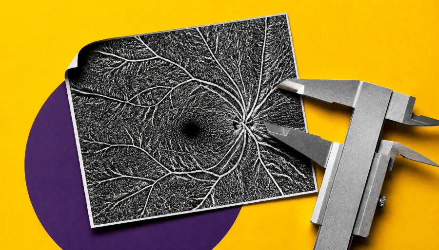
A new study finds that OCT angiography (OCT-A) biomarkers can detect small but meaningful changes in retinal blood vessels as diabetic retinopathy (DR) progresses. Researchers tracked 164 type 2 diabetes patients over three years, scanning eyes every four months. The findings suggest repeated OCT-A imaging could help clinicians monitor disease evolution and personalize patient care.
A new study published in Frontiers in Ophthalmology suggests that OCT angiography (OCT-A) could be a powerful tool for tracking how diabetic retinopathy (DR) progresses over time. Researchers from Vanderbilt University followed 164 patients with type 2 diabetes and 17 healthy controls, scanning 328 diabetic eyes every four months for three years — one of the most comprehensive longitudinal OCT-A studies to date.
The team analyzed seven microvascular metrics, including vessel density, foveal avascular zone area, and acircularity index. Eyes with severe nonproliferative DR showed greater annual losses in vessel and skeleton density, along with worsening vessel irregularity — signs of progressive ischemic damage. These trends were "small but statistically significant," pointing to gradual biological remodeling rather than measurement noise.
Key Takeaways:
Why it matters: For clinicians managing diabetic patients, OCT-A offers a noninvasive way to stratify risk, detect early progression, and potentially tailor treatment — moving toward a more personalized approach to DR management.