
Turn on cookies to sign in
Signing in saves your progress to your Curie account. We can only do that with cookies on — turn them on to continue.
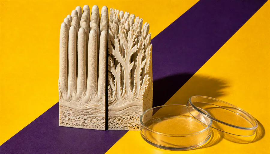
Your small intestine replaces its entire lining every few days — but how it stays so orderly has been a mystery. Yale researchers found that a cellular process called planar cell polarity (PCP), best known for guiding embryo development, is still active after birth and keeps intestinal cells crawling in the right direction. Disrupting PCP caused chaotic, branching villi structures in mice — shedding new light on gut architecture.
Your small intestine is a remarkably busy organ — it replaces its entire cellular lining every three to five days while maintaining a precise, organized structure that maximizes nutrient absorption. But how it pulls off this constant renewal without descending into chaos has puzzled scientists — until now.
Yale researchers publishing in Developmental Cell found that planar cell polarity (PCP) — a mechanism typically associated with fetal development — remains active in the small intestine after birth. PCP coordinates the direction in which cells orient and migrate, essentially acting like a traffic system that keeps intestinal cells crawling upward from the base (crypts) to the tips of finger-like structures called villi. When the team disrupted two PCP-regulating genes in mice, the crypts detached and floated toward the villus tips, and the villi branched abnormally instead of forming their characteristic finger-like shape.
The findings reframe how we understand intestinal cell migration — moving away from a passive "conveyor belt" model toward an active, coordinated crawling process guided by PCP.
Key Takeaways:
Why it matters: Understanding how the intestinal lining maintains its structure could open new avenues for research into conditions like inflammatory bowel disease, intestinal cancers, and malabsorption disorders — where this cellular organization breaks down.