
Turn on cookies to sign in
Signing in saves your progress to your Curie account. We can only do that with cookies on — turn them on to continue.
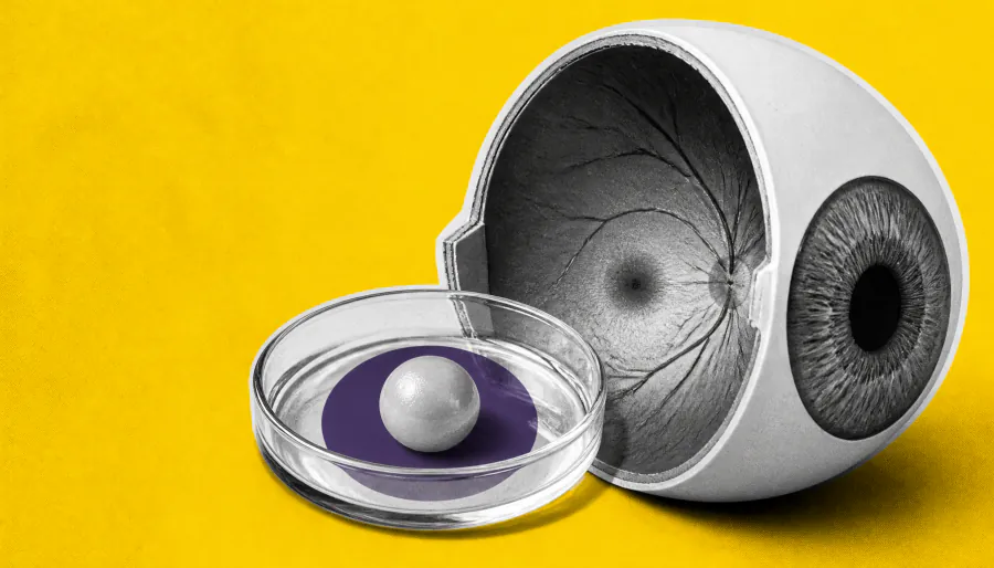
Johns Hopkins scientists have discovered that a vitamin A derivative and thyroid hormones work together to shape the foveola — the tiny retinal region responsible for our sharpest sight. Using lab-grown human retinal organoids, they found that early blue cone cells actually transform into red and green cones before birth, overturning a decades-old theory that those cells simply migrated away. The findings could one day guide cell replacement therapies for macular degeneration.
Johns Hopkins University researchers have upended a 30-year-old model of how human eyes develop sharp central vision. Using miniature human retinas grown in the lab — called organoids — the team discovered that a vitamin A derivative (retinoic acid) and thyroid hormones work in tandem to sculpt the foveola, the pinpoint region at the retina's center that powers detailed sight like reading and face recognition.
The big surprise? Blue cone cells that initially appear in the foveola during fetal development don't just migrate away — they appear to convert into red and green cone cells by week 14 of gestation. Retinoic acid first limits blue cone formation in the central retina, then thyroid hormones nudge the remaining blue cones to switch identity. This two-step process creates the precise cone arrangement essential for crisp central vision.
Key Takeaways:
Why it matters: Beyond developmental biology, this discovery opens a potential path toward cell replacement therapies for macular degeneration — a leading cause of central vision loss in older adults. The team aims to engineer precise populations of photoreceptors from organoids that could one day be transplanted to restore lost vision.