
Turn on cookies to sign in
Signing in saves your progress to your Curie account. We can only do that with cookies on — turn them on to continue.
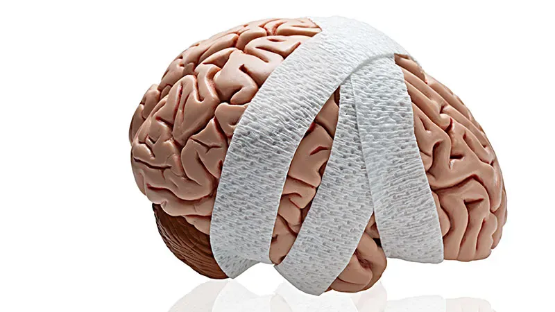
Early CT imaging could help predict which patients with nontraumatic acute brain injury will progress to brain death, a new meta-analysis finds. In stroke patients, signs of intracranial mass effect tripled the odds of death by neurologic criteria, while cardiac arrest patients who progressed to brain death showed notably lower gray-white matter ratios. The findings highlight CT as the most reproducible imaging tool for this critical prognosis.
A new meta-analysis published in BMC Neurology finds that early CT imaging markers can reliably predict progression to death by neurologic criteria (DNC) — commonly known as brain death — in patients with nontraumatic acute brain injury. The study analyzed 18 studies involving over 9,000 participants, focusing on conditions like hypoxic-ischemic encephalopathy, intracerebral hemorrhage, and ischemic stroke.
Researchers identified two key CT-based signals: in severe stroke patients, signs of intracranial mass effect (such as herniation or absent ambient cistern) were strongly linked to DNC, while in post-cardiac arrest patients, a lower gray-white matter ratio (GWR) — a marker of diffuse cerebral edema — was significantly associated with progression to brain death.
By the Numbers:
Why it matters: Predicting brain death early and accurately is critical for clinical decision-making, family communication, and organ donation planning. While evidence remains limited and largely retrospective, CT-based markers offer a promising, accessible tool for clinicians navigating these high-stakes situations.