

Turn on cookies to sign in
Signing in saves your progress to your Curie account. We can only do that with cookies on — turn them on to continue.
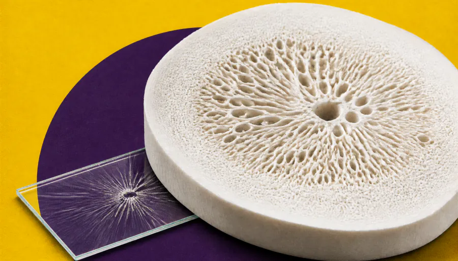
Scientists at Washington University have uncovered how a signaling protein called FGFR1 helps newly embedded bone cells survive. Without it, the surrounding bone matrix becomes disorganized and cellular communication networks break down, triggering cell death in newly formed bone. The findings could lay the groundwork for better understanding age-related bone fragility.
Scientists at Washington University School of Medicine have identified a critical role for the protein FGFR1 in keeping newly formed bone cells — called osteocytes — alive and functional. By selectively switching off the FGFR1 gene in adult mice, researchers found that the bone matrix surrounding newly embedded osteocytes became disorganized, and the microscopic communication network connecting these cells broke down, ultimately triggering cell death.
Importantly, the damage was confined to newly formed bone, not older, pre-existing bone. Dying cells still carried markers of immature bone-forming cells, suggesting FGFR1 is essential during the critical transition from bone-forming cell to fully mature osteocyte. Matrix disruption — including thinner collagen layers and disorganized fibers — appeared as early as three weeks after FGFR1 was inactivated, hinting that structural breakdown precedes cell death.
Key Takeaways:
Why it matters: Osteocyte loss is closely tied to brittle, fragile bones — a hallmark of aging. Understanding how these cells survive their earliest stages could open new avenues for research into osteoporosis and age-related bone disease.