Curie Brief
Turn on cookies to sign in
Signing in saves your progress to your Curie account. We can only do that with cookies on — turn them on to continue.
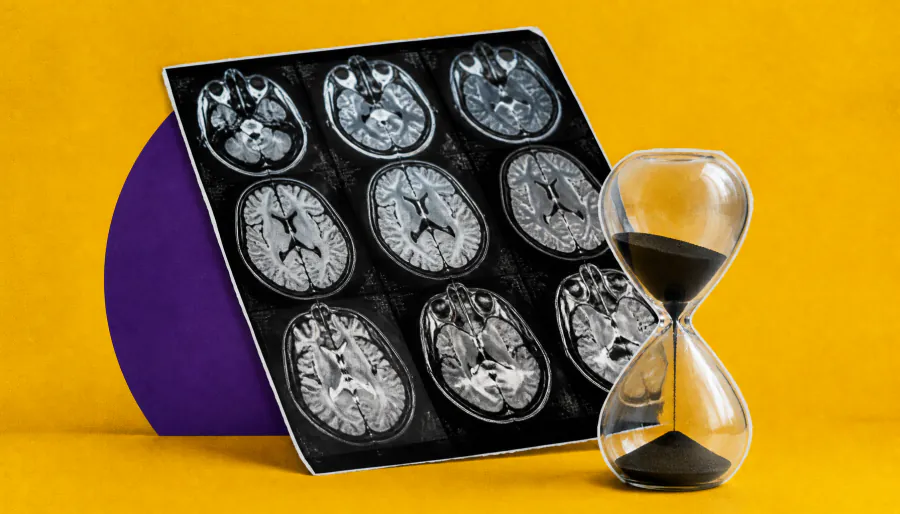
A new imaging measure could help detect frontotemporal dementia pathology while patients are still alive. Researchers found that Cortical Disarray Measurement (CDM), derived from diffusion MRI, corresponded with the microscopic distribution of brain pathology later confirmed at autopsy in 36 patients with behavioral variant frontotemporal dementia. The associations were strongest in cases with TDP-43 pathology.
A diffusion MRI–based tool may be able to detect the microscopic fingerprints of frontotemporal dementia (FTD) during a patient's lifetime — a potential game-changer for a disease that currently can only be definitively diagnosed after death. Researchers presented findings at the 15th International Conference on Frontotemporal Dementias showing that Cortical Disarray Measurement (CDM), developed by Oxford Brain Diagnostics, corresponded with autopsy-confirmed pathology patterns in patients with behavioral variant FTD (bvFTD).
The study analyzed 36 autopsy-confirmed cases of frontotemporal lobar degeneration (FTLD), comparing MRI measurements taken during life against postmortem pathology across 14 cortical regions. CDM's derived measure, PerpPD+, tracked closely with the laminar distribution of neuropathology — particularly in FTLD-TDP cases, where pathology tends to concentrate in superficial cortical layers.
Key Takeaways:
Why it matters: Frontotemporal dementia is notoriously difficult to diagnose and subtype during life. A non-invasive imaging biomarker that can reflect underlying molecular pathology could accelerate diagnosis, improve clinical trial enrollment, and ultimately open the door to more targeted treatments.