Curie Brief
Turn on cookies to sign in
Signing in saves your progress to your Curie account. We can only do that with cookies on — turn them on to continue.
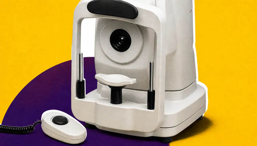
A new imaging tool may give eye doctors a head start on catching glaucoma before it worsens. OCT angiography (OCT-A) detected glaucoma progression earlier than standard OCT or visual field testing in a retrospective study — beating visual field tests by an average of 2.3 years. The catch? Its false-positive rate was higher, meaning it's best used alongside existing tools, not as a replacement.
A study published in JAMA Ophthalmology suggests that OCT angiography (OCT-A) may detect glaucoma progression earlier than the current gold standards — structural OCT and visual field testing. Researchers at the Shiley Eye Institute retrospectively analyzed 180 eyes from 116 glaucoma patients and found that OCT-A was the first to flag progression in the largest share of eyes, making it a potentially valuable addition to the monitoring toolkit.
That said, OCT-A isn't ready to fly solo. Its higher false-positive rate means clinicians need to interpret results carefully, and experts caution that the technology still lacks standardized progression criteria across devices.
By the Numbers:
Why it matters: Earlier detection of glaucoma progression could help clinicians intervene sooner and preserve patients' vision. But with a specificity of just 68.9%, OCT-A is best viewed as a complementary signal — especially when structural and functional tests disagree — rather than a standalone diagnostic tool.