Curie Brief
Turn on cookies to sign in
Signing in saves your progress to your Curie account. We can only do that with cookies on — turn them on to continue.
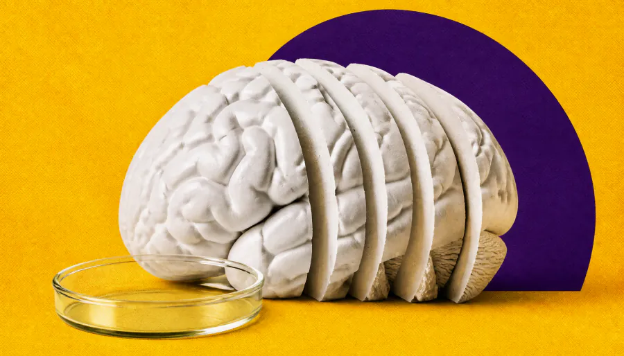
The brain just got a lot more complex. Researchers at EPFL have created the first detailed 3D map of lipids in the mouse brain, uncovering 539 distinct chemical zones called "lipizones." The atlas reveals hidden organizational patterns — including surprising complexity in white matter — and could open new doors for understanding Alzheimer's, depression, and other brain disorders.
Scientists at EPFL have published the first comprehensive 3D atlas of lipids — the fatty molecules that form neuron membranes, insulate nerve fibers, and facilitate cell communication — across the entire mouse brain. Published in Nature, the Lipid Brain Atlas reveals that these molecules follow a precise, highly organized architecture that both mirrors known brain anatomy and exposes chemical boundaries that traditional maps have missed entirely.
To build it, researchers used mass spectrometry imaging to analyze 172 lipids across 109 brain slices from 11 mice, generating ~7 million data points that were stitched into a 3D model using custom machine-learning tools. The result: 539 chemically distinct "lipizones," each with its own molecular fingerprint. Notably, white matter — long assumed to be chemically uniform — turned out to be a complex patchwork of distinct zones. The atlas also captured dramatic lipid shifts during pregnancy, with changes more pronounced than typical sex-based differences.
By the Numbers
Why it matters: Changes in brain lipids are increasingly linked to conditions like Alzheimer's disease and depression. This atlas gives researchers a healthy reference baseline to study where lipid organization breaks down — and the method, not just the map, could soon be applied to the human brain.