Curie Brief
Turn on cookies to sign in
Signing in saves your progress to your Curie account. We can only do that with cookies on — turn them on to continue.
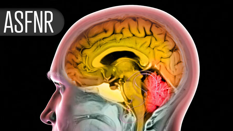
Even when a blood clot is successfully removed during stroke treatment, some patients still don't recover well — and a new imaging measure may explain why. A proof-of-concept study found that poor capillary bed perfusion, measured via post-thrombectomy perfusion MRI, predicted both infarct growth and poor 90-day functional outcomes better than conventional imaging parameters or baseline stroke severity.
Even after a successful thrombectomy — the gold-standard procedure for removing blood clots in acute ischemic stroke — a significant number of patients still end up with poor outcomes. A new proof-of-concept study presented at the American Society of Functional Neuroradiology meeting suggests that microvascular dysfunction, specifically poor capillary bed perfusion, may be the culprit hiding in plain sight.
Researchers at UCSF analyzed 47 stroke patients who had complete or near-complete reperfusion after thrombectomy. They found that a measure called relative transit time heterogeneity (RTH) — captured on perfusion MRI taken within 2 hours post-procedure — outperformed conventional imaging metrics like cerebral blood flow and volume in predicting both infarct progression and poor 90-day functional outcomes. Notably, 43% of patients experienced substantial infarct growth, and the same proportion had poor functional outcomes.
By the Numbers:
Why it matters: This research opens the door to identifying microvascular dysfunction as a potential treatment target for the "no-reflow" phenomenon in stroke care — a gap that current therapies don't address. A larger validation study is the critical next step.