Curie Brief
Turn on cookies to sign in
Signing in saves your progress to your Curie account. We can only do that with cookies on — turn them on to continue.
Spotting cancer cells in a sea of blood just got a whole lot smarter. Researchers developed a label-free platform that uses a spiral microfluidic chip to physically sort tumor cells by size, then deploys an AI model to identify them from standard microscope images — no fluorescent staining required. The system achieved nearly 90% tumor cell recovery and 96% identification accuracy.
Finding a handful of tumor cells among billions of blood cells is like searching for a needle in a haystack — and that's exactly what liquid biopsy tries to do. Researchers from Taiyuan University of Technology and Shanxi Bethune Hospital have developed a new platform that tackles this challenge without relying on molecular markers or fluorescent stains, making the process simpler and more flexible.
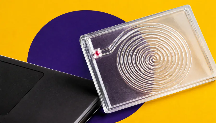
The system works in two steps. First, a spiral microfluidic chip uses the physical size difference between tumor cells (12–25 µm) and blood cells (6–12 µm) to sort them into separate flow paths. Then, a YOLOv8 deep-learning model analyzes standard brightfield microscope images to identify which collected cells are actually tumor cells — no extra labeling needed.
By the Numbers:
Why it matters: This proof-of-concept platform could streamline cancer monitoring through liquid biopsy, preserving cell integrity for downstream uses like single-cell sequencing and drug testing — potentially making early cancer detection faster and less invasive.