Curie Brief
Turn on cookies to sign in
Signing in saves your progress to your Curie account. We can only do that with cookies on — turn them on to continue.
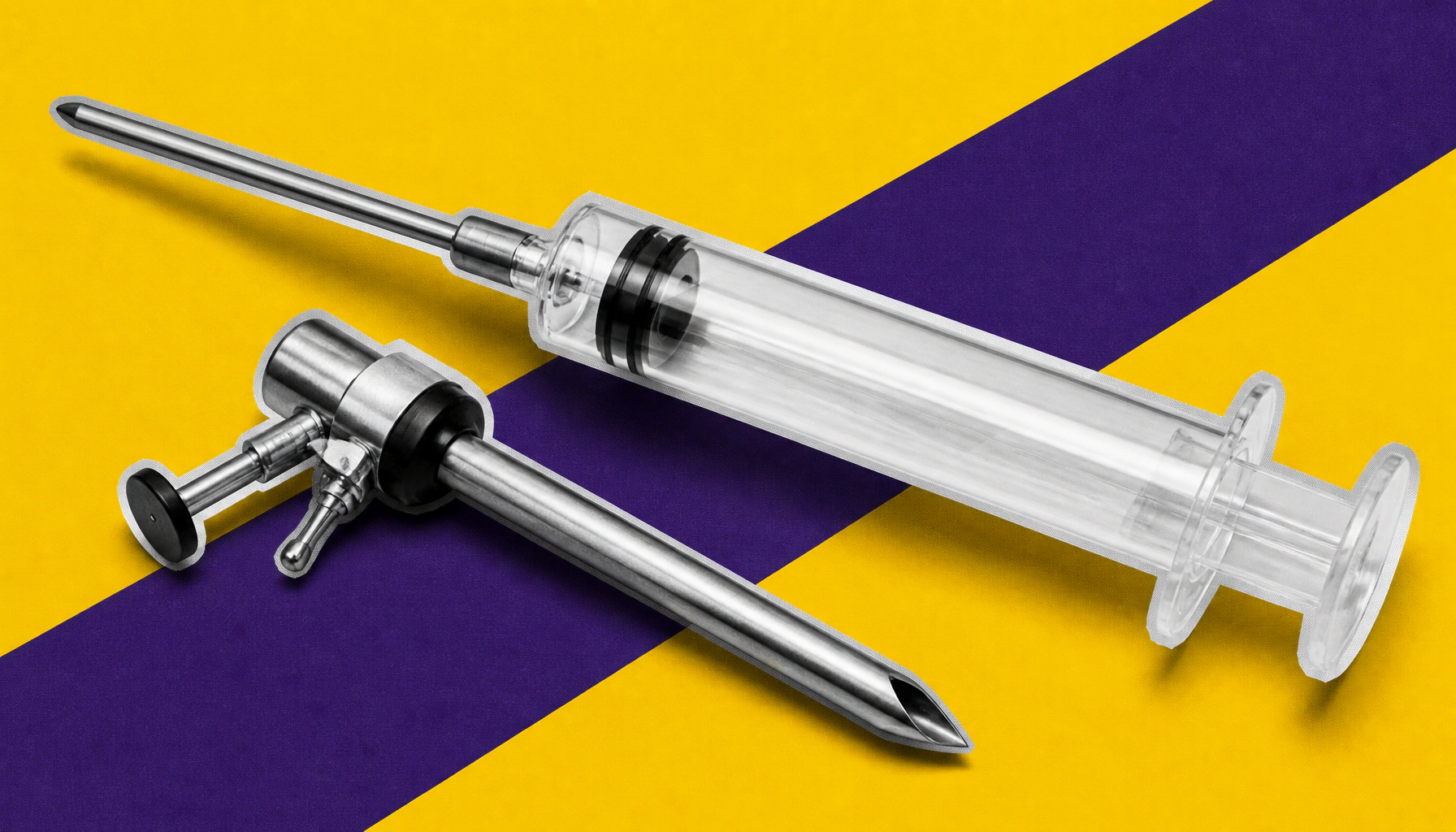
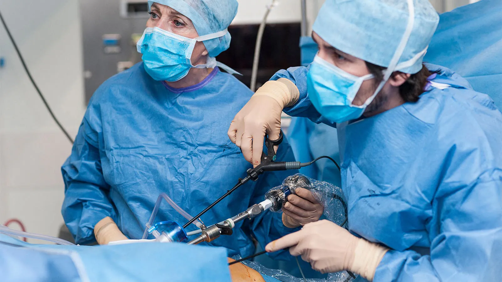
A new handheld device developed by MIT researchers could revolutionize early ovarian cancer detection by gently collecting living cells directly from fallopian tube tissue. The microfluidic tool uses fluid shear stress to harvest viable cells without destroying surrounding tissue, enabling diagnostics, disease modeling, and personalized treatment development. Early results show the cells remain alive and grow in culture far better than those collected by conventional methods.
Ovarian cancer is notoriously hard to catch early — and that's largely what makes it so deadly. Now, MIT engineers working with clinicians at Johns Hopkins have developed a handheld microfluidic device that could change that by collecting living cells directly from fallopian tube tissue, where most high-grade serous ovarian cancers originate.
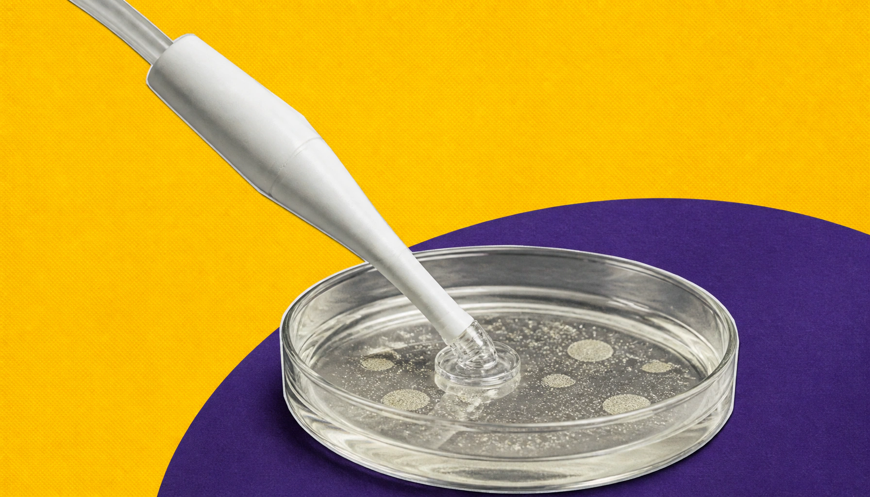
The device works by forming a vacuum seal against tissue and using fluid flow to apply gentle shear stress, detaching living cells from a precise, targeted area — without damaging the surrounding tissue. Those cells can then be grown into organoids, used for diagnostics, or tested against different treatments to guide personalized medicine. The team successfully collected viable cells from fresh human fallopian tube samples and grew them in culture, a feat that conventional chemical-preservation methods can't achieve.
By the Numbers:
Why it matters: This technology could eventually be integrated into routine pathology workflows or used to swab tissue inside living patients — opening the door to earlier, less invasive cancer detection across multiple cancer types, not just ovarian.