Curie Brief
Turn on cookies to sign in
Signing in saves your progress to your Curie account. We can only do that with cookies on — turn them on to continue.
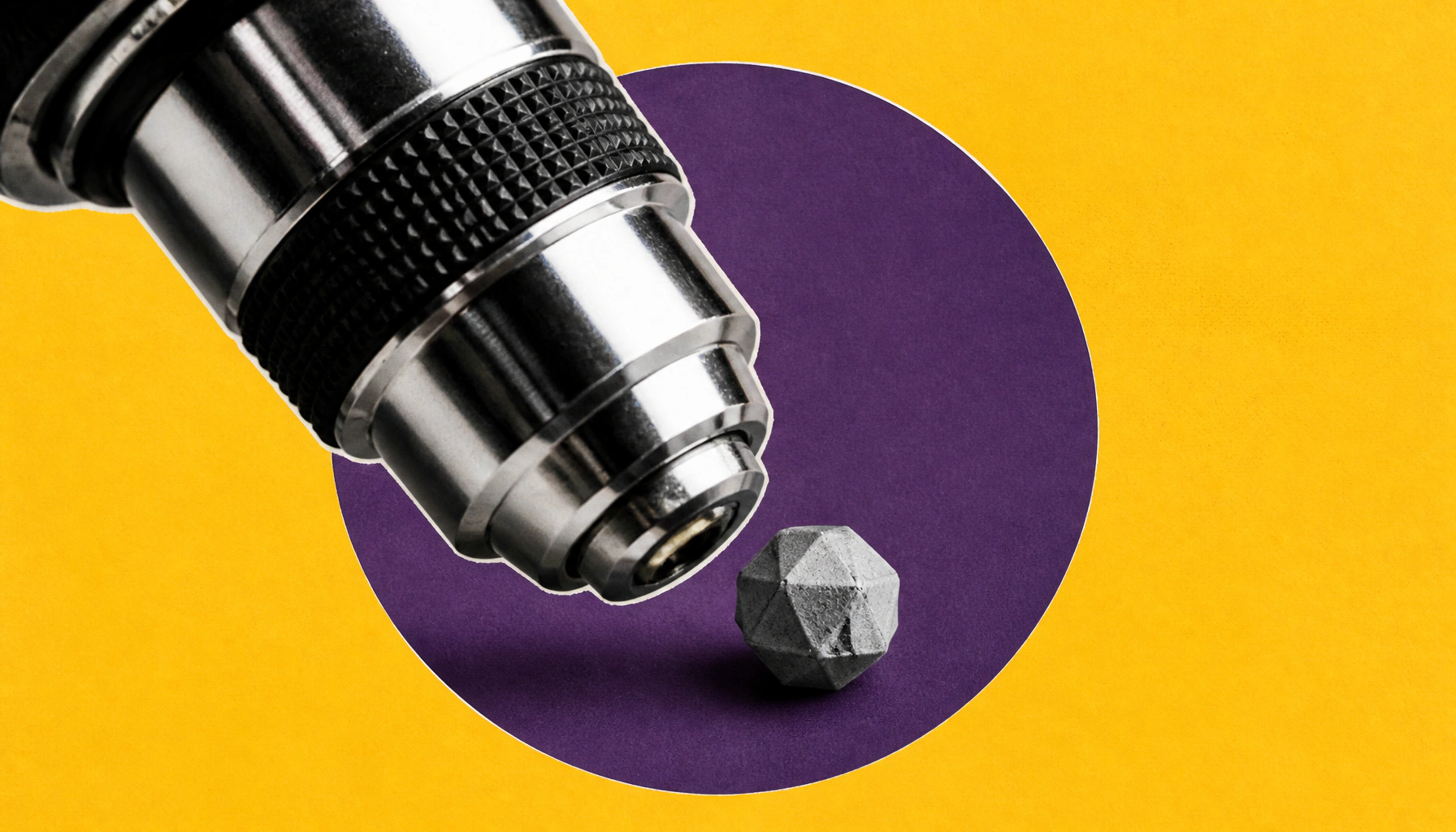
For the first time, Oxford researchers filmed the step-by-step self-assembly of individual virus-like particles, molecule by molecule. Using a technique called mass photometry, they revealed how viral protein shells reliably form despite thousands of possible configurations. The findings could open new doors for antiviral drug design and vaccine engineering.
For the first time, Oxford University researchers have captured live, real-time footage of individual virus-like particles assembling themselves — one protein at a time. Published in Nature, the study used mass photometry, a light-scattering technology developed at Oxford, combined with a new molecule-confinement method to "weigh" a growing particle repeatedly as new components joined it.
The team found that viral capsid assembly works like navigating a maze: protein building blocks initially form weak, reversible bonds — allowing failed arrangements to fall apart and retry. But when proteins lock into specific closed structures (starting with a stable pentagonal ring of five proteins), the process accelerates rapidly, funneling complexity into a reliable, step-by-step construction pathway.
Key Takeaways:
Why it matters: Understanding exactly how viruses build their protective shells — and where the process can be interrupted — gives scientists a precise molecular target for new antiviral therapies and smarter vaccine platforms.