Curie Brief
Turn on cookies to sign in
Signing in saves your progress to your Curie account. We can only do that with cookies on — turn them on to continue.
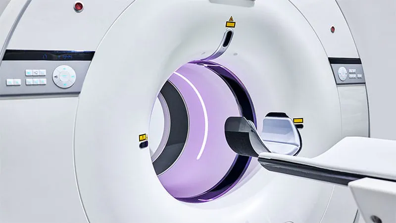
MRI scans taken during therapeutic hypothermia (cooling therapy) in newborns with hypoxic-ischemic encephalopathy (HIE) are just as reliable as scans taken nearly two weeks later. A study of 114 neonates found 95% concordance between early and later MRIs in identifying brain injury pattern and severity. This supports using early MRI to guide timely clinical decisions and family counseling.
When a newborn suffers from hypoxic-ischemic encephalopathy (HIE) — brain injury caused by oxygen deprivation around birth — doctors typically use therapeutic hypothermia (cooling the baby's body temperature) to limit damage. But knowing how bad the injury is, and when to image it, has been a clinical challenge. A new study suggests the answer may come sooner than previously thought.
Researchers at McGill University retrospectively analyzed 114 newborns with HIE who underwent therapeutic hypothermia between 2008 and 2023. They compared MRI scans taken during the cooling period (median age: 2 days) with scans taken after cooling ended (median age: 11 days). The results were striking: the two sets of scans agreed 95% of the time on both the pattern and severity of brain injury.
By the Numbers:
Why it matters: Getting an accurate read on brain injury early could transform care for these critically ill newborns — enabling faster prognostication, earlier family counseling, and quicker enrollment in clinical trials. While follow-up MRI is still recommended in complex cases, this study makes a strong case for broader use of early imaging in well-equipped centers.