
Turn on cookies to sign in
Signing in saves your progress to your Curie account. We can only do that with cookies on — turn them on to continue.
Keratoconus is routinely diagnosed years after symptoms first appear — but it doesn't have to be. Integrating advanced corneal imaging into standard pretesting workflows can help eye care providers catch the disease earlier, before vision loss becomes significant. With a new FDA-approved cross-linking procedure now available for patients as young as 13, early detection has never been more actionable.
Keratoconus — a progressive corneal disorder that distorts vision — is frequently diagnosed years too late. A prospective multicenter study of 421 patients found that symptoms preceded diagnosis by nearly three years on average, with many patients not presenting until they already have moderate or severe disease in their 30s. Traditional tools like slit-lamp exams and autokeratometry simply aren't sensitive enough to catch early-stage disease.
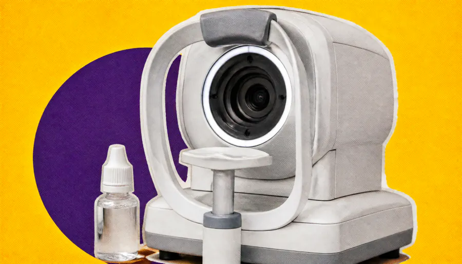
The fix, according to OD John Gelles, is straightforward: swap out autorefraction/autokeratometry for combination devices that pair topography or tomography with autorefraction. These tools map the full corneal contour and can be used in roughly the same time as conventional pretesting — making keratoconus screening a seamless part of routine comprehensive eye care rather than a separate specialty workflow.
Key Takeaways:
Why it matters: The FDA recently approved epithelium-on cross-linking (Epioxa) for patients aged 13 and up — labeled for keratoconus broadly, not just progressive cases. Earlier detection means more patients can access this vision-preserving treatment before significant damage occurs.