
Turn on cookies to sign in
Signing in saves your progress to your Curie account. We can only do that with cookies on — turn them on to continue.
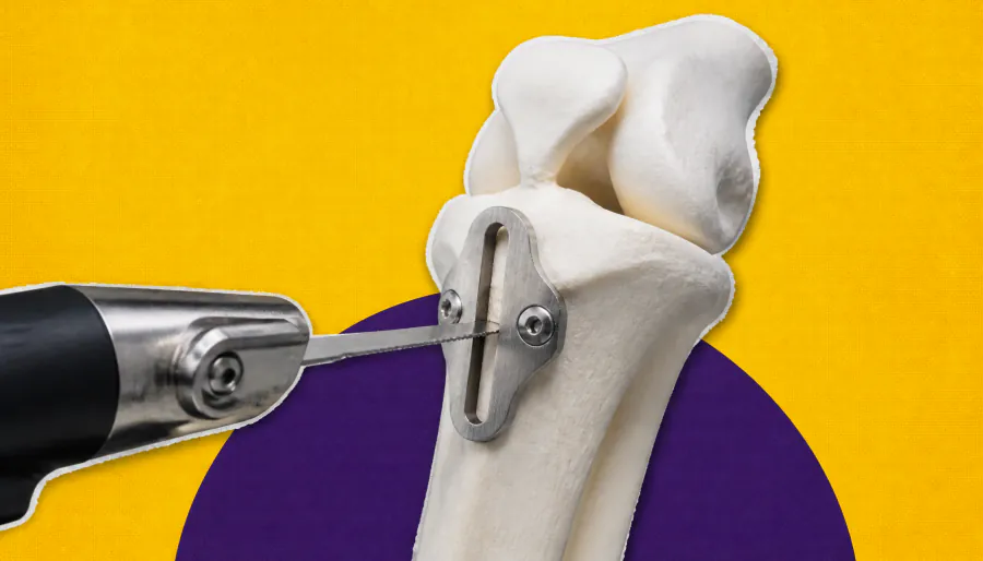
Precision planning is reshaping how surgeons approach tibial tubercle osteotomy (TTO) for patellar instability. Yale orthopedic surgeon Dr. John Fulkerson outlines how 3D CT imaging and personalized surgical cutting guides now allow surgeons to tailor the exact direction and degree of bone repositioning for each patient — improving outcomes for conditions like recurrent patellar dislocation and patella alta.
Tibial tubercle osteotomy (TTO) — a procedure that repositions the bony attachment of the kneecap tendon — is a cornerstone treatment for patellar instability, but it's far from one-size-fits-all. Yale orthopedic surgeon Dr. John Fulkerson argues that the real challenge isn't just deciding whether to perform a TTO, but determining exactly how to move the tubercle — and by how much — for each individual patient.
Common indications include lateral patellofemoral arthritis, patellar maltracking, patella alta, and recurrent patellar dislocation. The most frequently performed variant, the anteromedial TTO, unloads the joint, corrects alignment, and improves patellar engagement with the trochlear groove. Adding distalization can further optimize patella positioning for patients with patella alta.
Key Takeaways:
Why it matters: As orthopedic surgery embraces personalized medicine, custom-guided TTO offers the potential to reduce surgical variability, improve functional outcomes, and minimize complications in patients with complex patellar instability.