Curie Brief
Turn on cookies to sign in
Signing in saves your progress to your Curie account. We can only do that with cookies on — turn them on to continue.
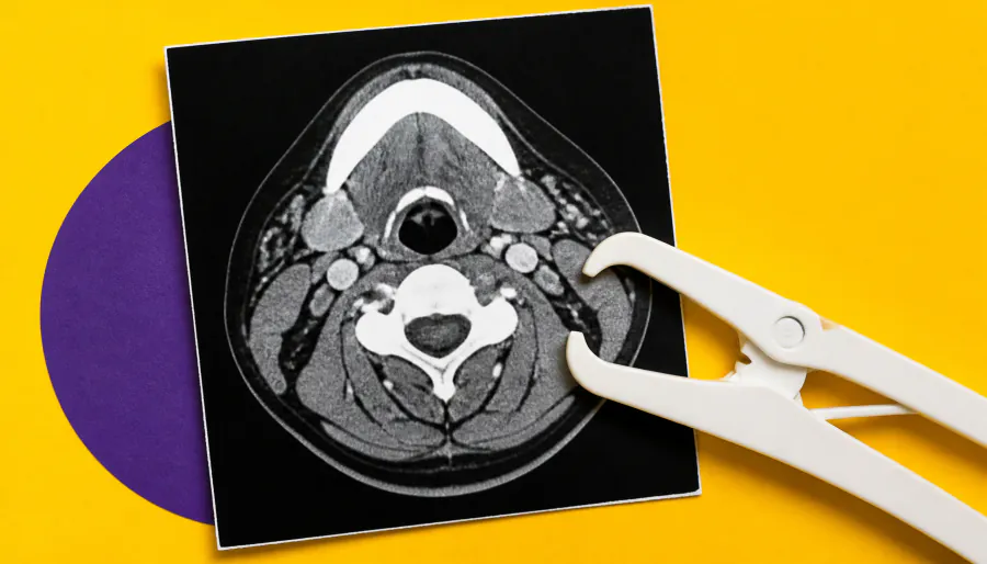
More muscle, better odds. A new study found that head and neck cancer patients with greater skeletal muscle mass on pre-treatment CT scans had significantly improved overall survival after radiotherapy. Conversely, higher intermuscular fat was linked to worse outcomes. Researchers say integrating this automated body composition analysis into clinical workflows could sharpen risk stratification for vulnerable patients.
A new study published in JAMA Otolaryngology–Head & Neck Surgery found that body composition — specifically skeletal muscle mass and intermuscular fat — is a meaningful predictor of survival in head and neck cancer patients undergoing radiotherapy (RT). Researchers at UCLA developed a deep-learning tool to automatically quantify 3D cervical neck soft tissue volumes from routine pre-treatment CT scans, removing the need for manual analysis.
The retrospective cohort study analyzed 659 patients treated with definitive RT between 2014 and 2024, with a median follow-up of 5.5 years. The tool revealed striking variation in tissue composition across BMI categories — and showed that BMI alone doesn't tell the whole story.
By the Numbers:
Why it matters: BMI is a blunt instrument — this AI-powered approach goes deeper, capturing muscle quality and fat distribution that standard metrics miss. Integrating this opportunistic phenotyping into clinical workflows could help clinicians flag high-risk patients earlier and tailor supportive care to help them endure cancer-directed therapy.