Curie Brief
Turn on cookies to sign in
Signing in saves your progress to your Curie account. We can only do that with cookies on — turn them on to continue.
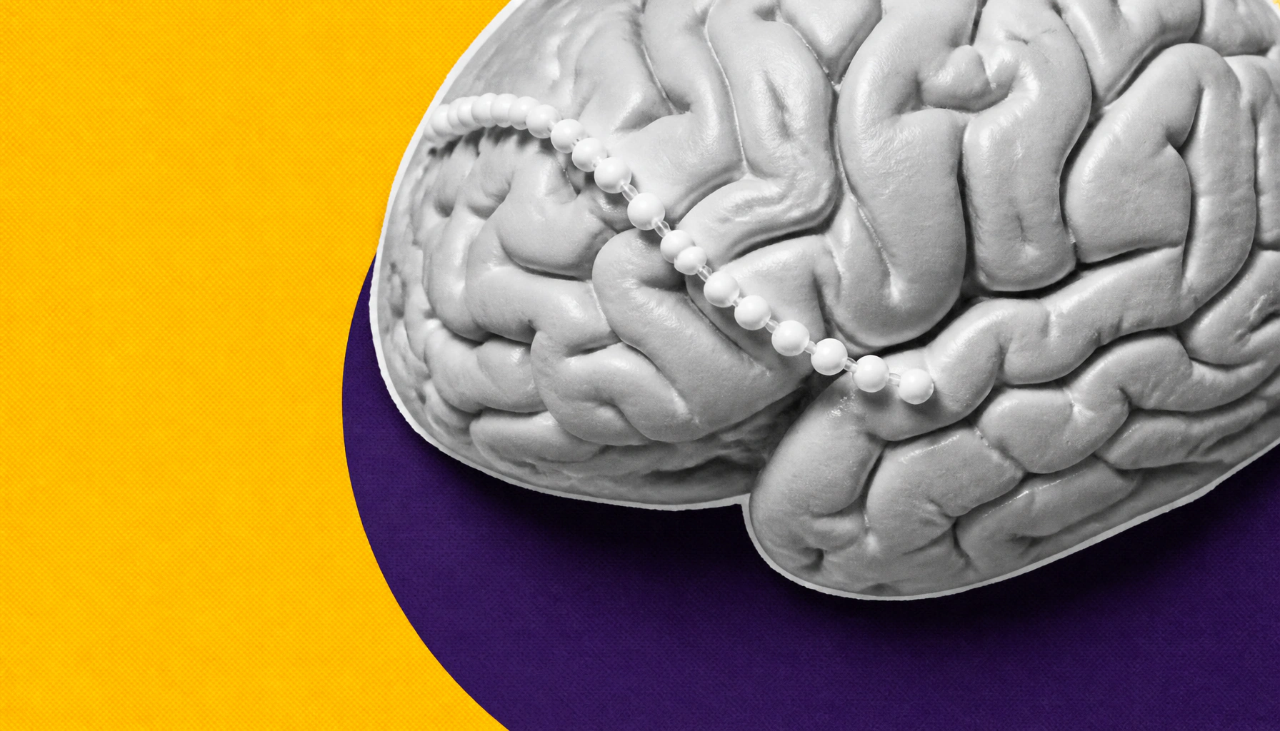
Neuroscience textbooks may have gotten axon anatomy wrong for over a century. Johns Hopkins researchers found that brain cell axons naturally resemble strings of tiny pearls — not smooth tubes — and these pearl-like structures dynamically shift with neural activity, influencing how fast electrical signals travel. The findings have since been confirmed in human brain tissue too.
For more than 100 years, neuroscience textbooks have depicted brain cell axons as smooth, narrow tubes. But researchers at Johns Hopkins Medicine say that picture may be fundamentally wrong. Using high-pressure freezing electron microscopy — a technique that preserves tissue closer to its natural state — they found that axons in mouse neurons are studded with repeating, nanoscale bulges, making them look more like strings of pearls. Importantly, these aren't signs of damage; they appear to be a normal feature of axon architecture.
The pearl-like structures aren't just cosmetic. Their shape influences how quickly electrical signals move through the brain, and they're not static — after high-frequency electrical stimulation, the pearls grew an average of 8% longer and 17% wider, with signal-slowing effects lasting over an hour. Cholesterol in the membrane also plays a key role in maintaining the structure and signaling speed. A follow-up study published in Neuron confirmed pearled axons in human cortical tissue, extending the findings beyond mice.
Key Takeaways:
Why it matters: If axon structure is more dynamic and complex than previously thought, it could reshape our understanding of how the brain learns, adapts, and breaks down in disease — with potential implications for neurological and neurodegenerative conditions.