Loading Curie Briefs...
Getting the latest healthcare news for you
Getting the latest healthcare news for you

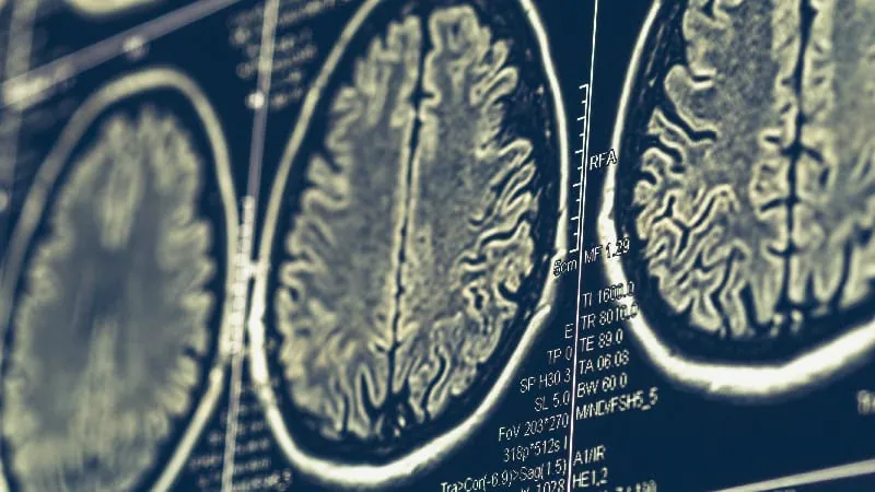
An MRI-based measure of cortical atrophy can predict how quickly patients with early-onset Alzheimer's disease (EOAD) will progress from mild cognitive impairment to dementia. A study of 130 patients found that greater baseline atrophy in a specific brain signature region raised the risk of progression by 24% per standard deviation increase. This low-cost tool could help personalize care and improve clinical trial enrollment.
Researchers have found that a routine MRI scan can help predict how quickly patients with early-onset Alzheimer's disease (EOAD) will progress from mild cognitive impairment (MCI) to full dementia — potentially transforming how clinicians plan care for younger patients. The study, published in Neurology, followed 130 patients aged 40–64 with biomarker-confirmed EOAD over an average of nearly 22 months.
The key finding: greater baseline cortical atrophy within a specific EOAD-signature brain region — predominantly parietotemporal areas — was a strong predictor of faster disease progression. Notably, hippocampal volume, a commonly used marker, did not predict progression, suggesting that the EOAD-signature region is a more sensitive tool for this population.
By the Numbers
Why it matters: Early-onset Alzheimer's affects people during their most productive years, making accurate prognosis critical for patients, families, and clinical trial planning. A widely available, cost-effective MRI biomarker that can individualize risk prediction could be a game-changer for earlier intervention and treatment enrollment.