Loading Curie Briefs...
Getting the latest healthcare news for you
Getting the latest healthcare news for you

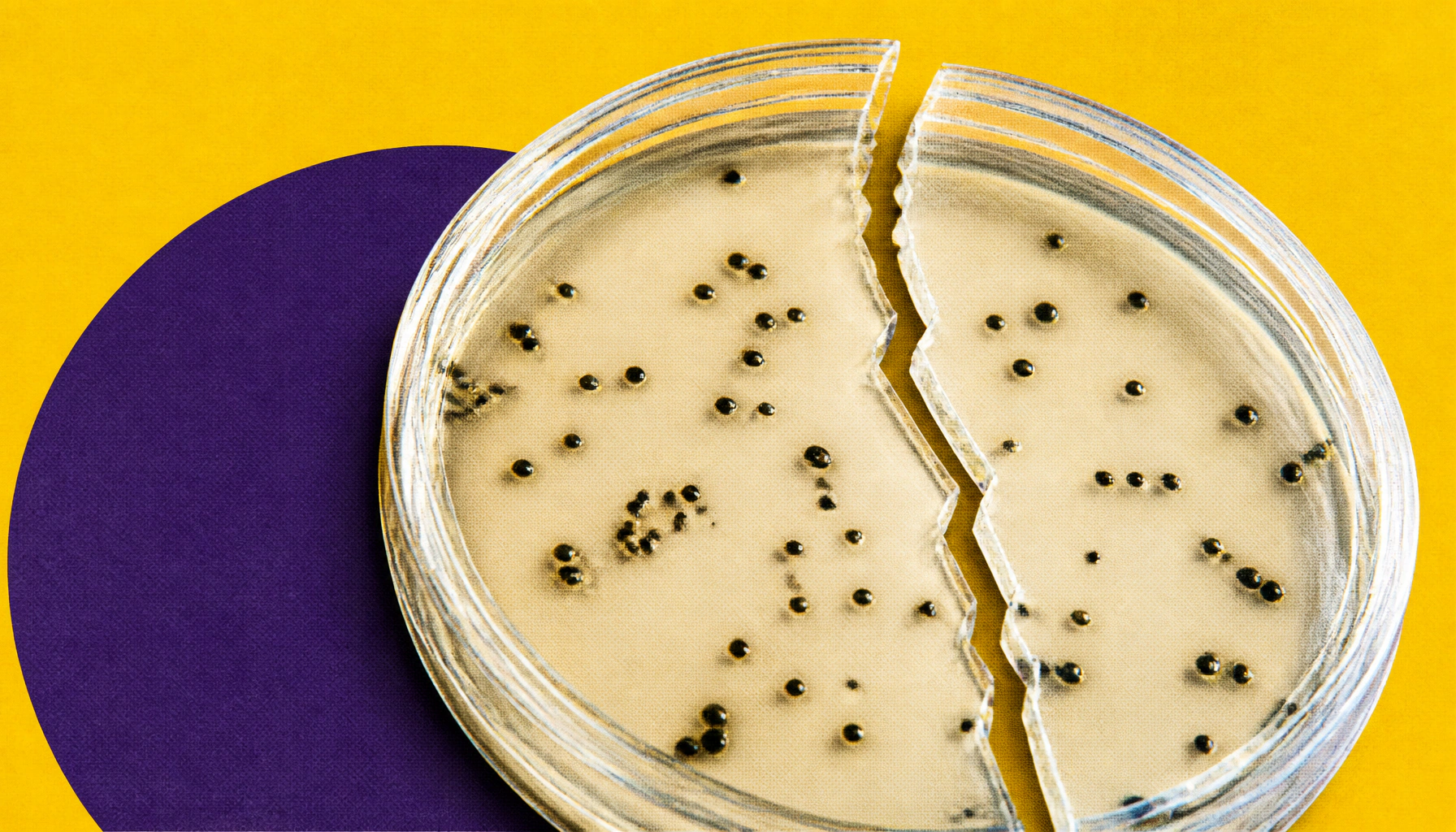
Researchers have captured, for the first time, how bacteria assemble their antibiotic-resistant outer shells. Using cryo-electron microscopy, a Japanese-led team revealed four structural snapshots of a key protein handoff process that helps Gram-negative bacteria build their protective outer membrane. The findings could open new doors for developing drugs that dismantle this bacterial armor.
Researchers at Japan's Nara Institute of Science and Technology (NAIST) have uncovered a critical step in how Gram-negative bacteria — notorious for their resistance to antibiotics — construct their protective outer membrane. Using cryo-electron microscopy (cryo-EM), the team captured four distinct structural snapshots of the SurA–BAM complex, the molecular machinery responsible for folding and inserting proteins into the bacterial outer membrane.
The study, set to publish in Nature Communications, shows that the chaperone protein SurA undergoes dramatic shape-shifting as it delivers unfolded outer membrane proteins (OMPs) to the BAM assembly complex. Key interactions — particularly between SurA's flexible P2 domain and a BAM component called BamE — help position SurA close enough to hand off its cargo. When researchers disrupted this interaction, OMP assembly dropped significantly.
Key Takeaways:
Why it matters: Antibiotic resistance is one of the most urgent global health threats. By mapping exactly how bacteria build their protective barriers, this research lays the groundwork for a new class of antibacterial agents designed to weaken — or collapse — that defense.