Loading Curie Briefs...
Getting the latest healthcare news for you
Getting the latest healthcare news for you

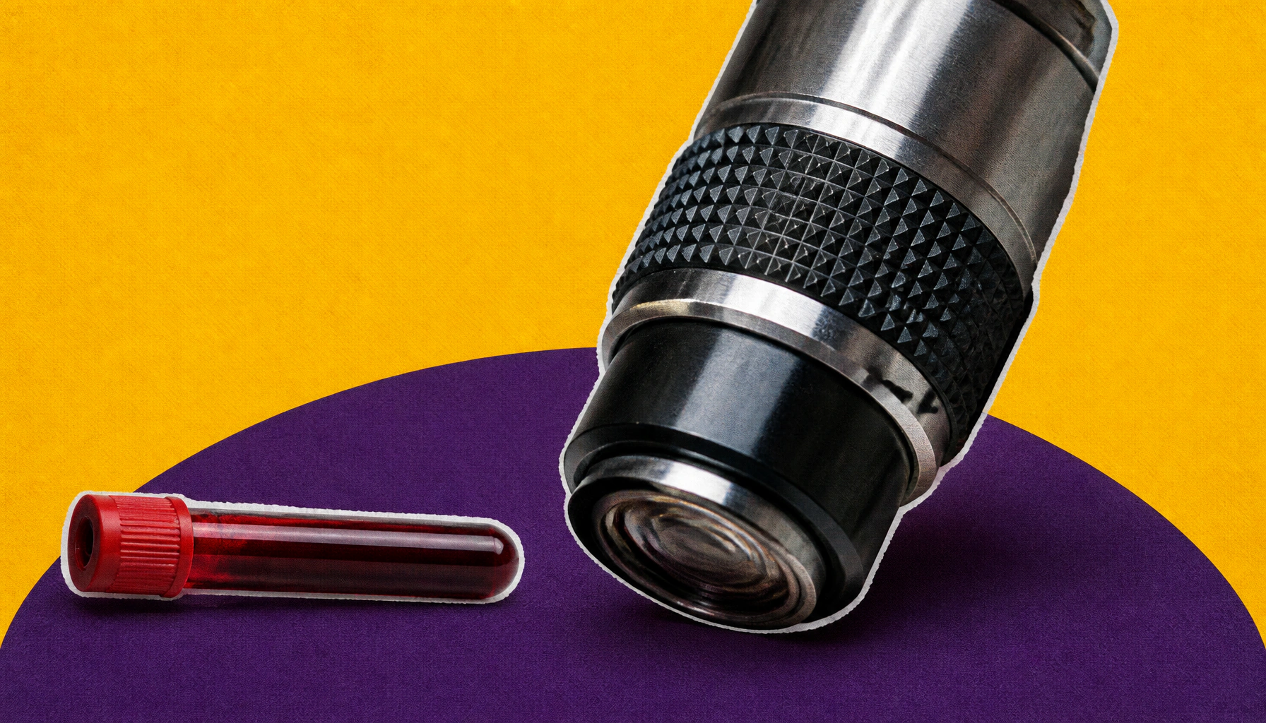
Researchers have developed a nondestructive imaging method that can assess the metabolic state of individual immune cells — without harming them. Using optical metabolic imaging (OMI), the team accurately identified immune cell activity in complex blood samples, opening the door to better diagnostics and smarter cell therapies like CAR T.
Scientists at the Morgridge Institute for Research have developed a new way to study immune cells that doesn't require destroying them in the process. Using a technique called optical metabolic imaging (OMI), the team can now assess the metabolic activity of single immune cells within complex blood samples — a first for this type of nondestructive analysis. The approach uses two long-wavelength photons to excite the natural fluorescence of metabolic byproducts in cells, revealing whether they're active or dormant.
The implications are significant for both diagnostics and treatment. Because the cells remain intact after imaging, they can still be used downstream — for example, to assess the fitness of immune cells before they're engineered into CAR T cell therapies. This could help clinicians make smarter decisions about which cells to use and improve patient outcomes.
By the Numbers
Why it matters: Current clinical tools give a basic count of immune cells but miss nuanced information about how those cells are functioning. OMI adds a metabolic layer to that picture — potentially transforming how conditions like blood cancers, lupus, and sepsis are diagnosed and treated.