Loading Curie Briefs...
Getting the latest healthcare news for you
Getting the latest healthcare news for you

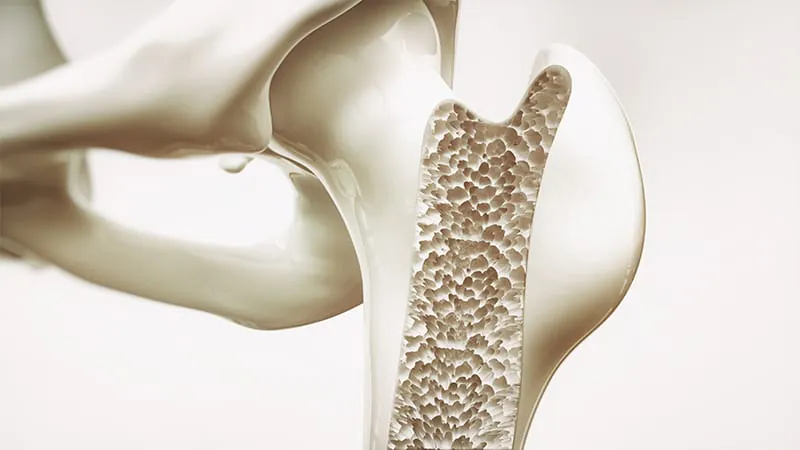
When it comes to catching osteoporosis and osteopenia early, lumbar CT scans are pulling ahead of MRI. A large meta-analysis of 42 studies found that CT-derived Hounsfield unit (HU) measurements consistently outperformed MRI-based bone quality scores in diagnostic accuracy. The findings suggest CT could be seamlessly woven into routine imaging workflows for opportunistic bone density screening.
When it comes to catching osteoporosis and osteopenia early, lumbar CT scans are pulling ahead of MRI. A systematic review and meta-analysis of 42 studies involving over 9,200 participants found that CT-derived Hounsfield unit (HU) measurements consistently outperformed MRI-derived vertebral bone quality (VBQ) scores across multiple diagnostic metrics — including accuracy, sensitivity, and specificity.
Complementing this, a separate retrospective study found that automated bone density extraction from thoracic CT scans is also highly reproducible across different scanners and imaging protocols, further strengthening the case for opportunistic osteoporosis screening via routine CT.
Together, these findings point to a compelling opportunity: patients already undergoing CT scans — whether for lung cancer screening or other reasons — could simultaneously be assessed for bone health, helping close a significant osteoporosis treatment gap.
By the Numbers:
Why it matters: Osteoporosis is vastly underdiagnosed. These findings support integrating HU measurements into routine CT workflows — turning scans patients are already getting into a powerful, no-added-cost screening tool for bone disease.