Loading Curie Briefs...
Getting the latest healthcare news for you
Getting the latest healthcare news for you

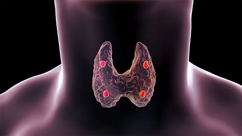
When first-line imaging fails, 4D CT delivers. A retrospective study of 73 patients with primary hyperparathyroidism found that four-dimensional CT reliably localized and lateralized parathyroid adenomas after inconclusive nuclear medicine and ultrasound scans. With high specificity and strong positive likelihood ratios, it helped avoid extensive four-quadrant surgical exploration in roughly 70% of cases, enabling a more targeted, minimally invasive approach.
When standard imaging hits a dead end, 4D CT may be the answer. A retrospective study published in the British Journal of Radiology evaluated multiphase (four-dimensional) CT as a second-line imaging tool in 73 patients with primary hyperparathyroidism whose initial nuclear medicine scans (99mTc-sestamibi SPECT/CT) and ultrasound yielded negative or conflicting results.
The study divided patients into a "negative" cohort (no adenoma found on any first-line scan) and a "discordant" cohort (contradictory or inconclusive findings). 4D CT used a three-phase contrast protocol to capture arterial and delayed enhancement patterns characteristic of parathyroid adenomas.
Key Takeaways:
Why it matters: For surgeons and endocrinologists, 4D CT offers a practical next step when first-line imaging is inconclusive — improving surgical planning and reducing the need for more extensive operations in patients with primary hyperparathyroidism.