Loading Curie Briefs...
Getting the latest healthcare news for you
Getting the latest healthcare news for you

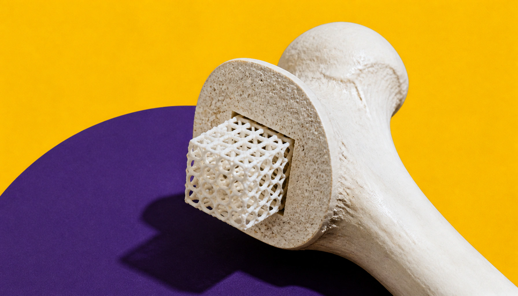
Penn State researchers have used genetic "switches" — tiny microRNA molecules — to program 3D-bioprinted stem cell clusters to regenerate bone tissue and grow new blood vessels. In mouse models, the bioprinted spheroids dramatically boosted bone healing compared to untreated controls. The findings could one day help patients who've lost bone due to severe trauma, cancer, or infection.
A Penn State interdisciplinary team has taken a major step toward using 3D bioprinting to rebuild bone — and the secret ingredient is genetic programming. By introducing two specific strands of microRNA (miR-148b and miR-210) into undifferentiated stem cells, researchers nudged the cells toward either bone growth or vascularization (the formation of new blood vessels) before assembling them into tiny cell clusters called spheroids. These spheroids were then precisely placed into scaffolds using a technique called aspiration-assisted bioprinting, which allows researchers to control the exact positioning of each cluster.
The results, published in Chemical Engineering Journal, are promising. In mouse models with bone tissue damage, untreated mice saw about 35% bone regeneration after six weeks, while mice treated with the bioprinted scaffold alone reached 93% coverage. Combining both microRNA types in the scaffold led to even better bone development and vascularization, with higher expression of CD31 — a key marker for blood vessel formation.
By the Numbers
Why it matters: Bone loss from trauma, cancer, or infection is notoriously hard to treat. This research lays the scientific groundwork for a future therapy that could rebuild complex bone structures — blood vessels and all — using programmed stem cells.