A surprisingly named imaging preset is changing how doctors see through knee implants. A new study found that the "Dental" metal artefact reduction setting on photon-counting CT angiography dramatically cuts through the image distortion caused by total knee replacements. This means clearer views of the blood vessels around the knee — a big win for vascular assessment in joint replacement patients.
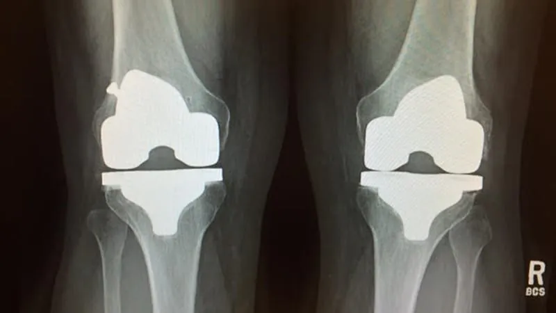
When it comes to imaging patients with total knee replacements, metal implants have long been the enemy of clarity — distorting CT scans and making it hard to assess nearby blood vessels. A new observational study suggests a counterintuitive fix: a reconstruction preset originally designed for dental imaging.
Researchers at University Hospital Würzburg analyzed ultra-high-resolution photon-counting CT angiography (PCCTA) scans from 25 patients with knee replacements, comparing eight different image reconstruction approaches. The "Dental" iterative metal artefact reduction (IMARdental) preset came out on top — by a wide margin — for both objective image quality and clinicians' subjective preference for assessing the popliteal artery (the key blood vessel behind the knee).
By the Numbers:
Why it matters: As knee replacements become increasingly common in aging populations, the ability to accurately image surrounding vasculature is critical for diagnosing complications. This study offers a practical, immediately applicable imaging tweak that could improve diagnostic confidence without any new equipment.