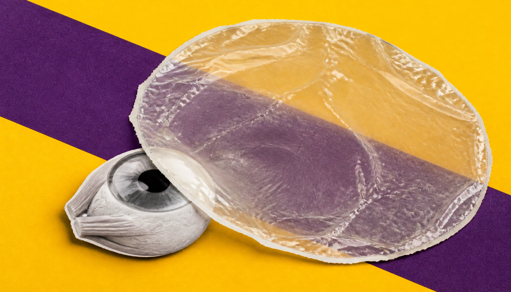
Amniotic membrane transplantation (AMT) has become one of ophthalmology's most versatile tools for treating a wide range of ocular surface diseases. Derived from the innermost layer of the placenta, the membrane offers anti-inflammatory, anti-scarring, and healing-promoting properties. From corneal ulcers to chemical burns and pterygium surgery, AMT is now a routine part of ocular surface reconstruction.
Amniotic membrane transplantation (AMT) has quietly become one of ophthalmology's most powerful biological tools. First used in eye care in the 1940s and reintroduced in the 1990s, AMT leverages the unique properties of the placenta's innermost layer — a thin, avascular membrane packed with biological activity — to restore damaged ocular surfaces.
The membrane works on multiple fronts: it acts as a scaffold for cell migration, suppresses inflammation by trapping inflammatory cells and reducing pro-inflammatory cytokines, inhibits scar formation, blocks abnormal blood vessel growth, and even has mild antimicrobial properties. It can be applied as an inlay graft (integrated into tissue defects), an overlay patch (acting like a biological bandage), or in a sandwich technique for deeper injuries.
Key Takeaways:
Why it matters: As ocular surface disease affects millions globally, AMT offers a biologically rich, relatively accessible solution that addresses inflammation, scarring, and healing simultaneously — making it a cornerstone of modern ophthalmic surgery.