Diagnosing motor neuron disease (MND) just got a potential upgrade. Researchers at Manchester Metropolitan University are combining ultrasound imaging with AI to detect MND non-invasively — potentially reducing the need for repeated, painful needle electromyography exams. Early results show diagnostic accuracy above 85%, and the team is now working to test the technology in patients at the earliest stages of disease.
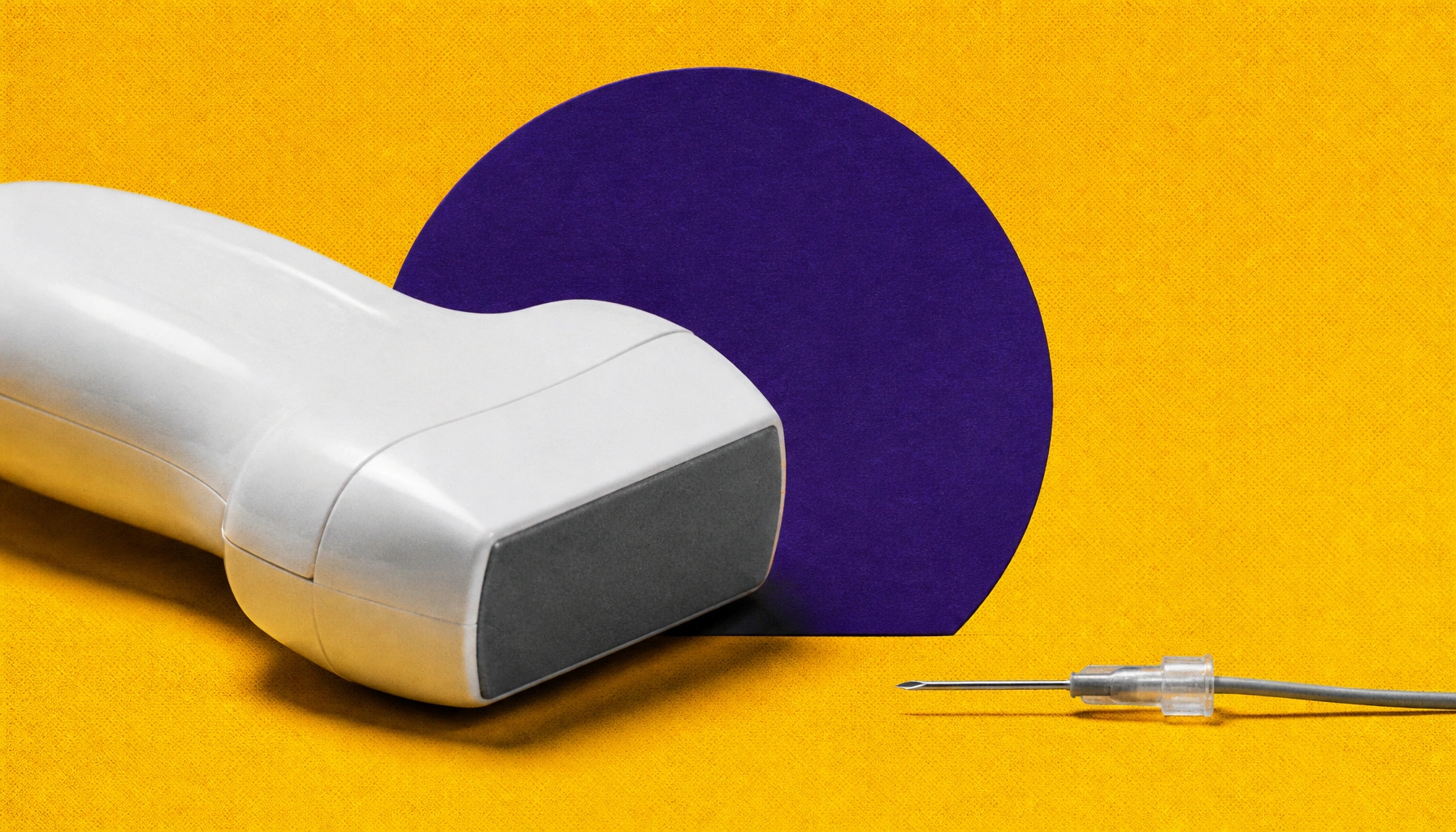
Diagnosing motor neuron disease could soon be a lot less painful. Professor Emma Hodson-Tole and her team at Manchester Metropolitan University are developing an AI-powered ultrasound tool designed to detect motor neuron disease (MND) without the need for invasive needle electromyography (EMG) — currently the gold-standard but notoriously uncomfortable diagnostic test. The technology uses computer vision to analyze muscle movement in ultrasound videos, identifying involuntary muscle twitches (fasciculations) and changes in muscle size and architecture that are hallmarks of MND.
The goal isn't to replace needle EMG entirely, but to give clinicians an additional, pain-free tool that can help determine whether invasive testing is even necessary — and potentially flag the disease earlier, when intervention matters most. The team is also prioritizing explainability, ensuring clinicians can understand why the AI reaches a given conclusion, not just what it concludes.
By the Numbers:
Why it matters: Earlier MND diagnosis means faster access to clinical trials and experimental therapies that may slow progression. A non-invasive, scalable tool could meaningfully transform how this devastating disease is detected and monitored in routine clinical care.